This is an education resource only. Ordering of all procedure codes on this website are subject to the Canberra Health Services guidelines for imaging orders.

INDICATIONS (1-5)
-
Ischaemic stroke to diagnose occlusion and thrombosis / assess change and monitor known cases
-
Carotid artery dissection
-
Vertebral artery dissection
-
Arterial vascular malformation within the head and neck
-
Transient ischaemic attack, used to evaluate carotid artery stenosis as potential causation
-
Subarachnoid haemorrhage to assess aneurysmal origin
-
Cerebral parenchymal haemorrhage, to assess for the presence of a vascular malformation or ongoing bleeding (spot sign)
-
Vascular trauma
-
Suspected vasculitis/fybromuscular dysplasia (FMD)/reversible cerebral vasoconstriction syndrome (RCVS)
PATHOLOGY DEMONSTRATED (6)
-
Occlusion and thrombosis within the carotid and vertebral arteries
-
Carotid artery dissection
-
Vertebral artery dissection
-
Arteriovenous malformation within the head and neck
-
Carotid artery stenosis
-
Aneurysms within cerebral and neck vasculature
-
Acute haemorrhage (spot sign)
PATIENT PREPARATION
-
Patient able to lie still for ten minutes
-
Not claustrophobic (sedation may be given)
-
Cognitively capable of following basic instructions
-
Metal artefacts removed from the region of interest
-
No respiratory distress when lying supine
-
Not allergic to Iodine based Contrast
-
No known kidney disease (eGFR below 30 as per RANZCR), however, acute setting consultant may sign to continue with poor renal function
-
No hyperthyroidism, may induce thyroid storm
-
Patient to have 18G cannula in anterior cubital fossa to enable a 7ml/s flow rate (20G cannula acceptable if flushing to 5ml/s).
-
Preferably patient fasted for 4 hours
ANATOMY INCLUDED

CT Brain - Non Contrast (Axial)
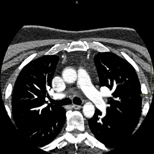
Angiography Arch to COW (Axial)

CT Brain - Non Contrast (Coronal)
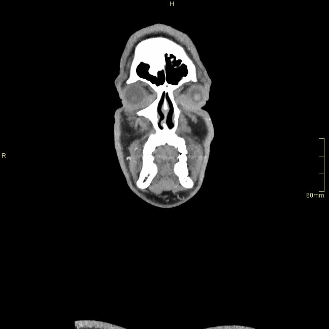
Angiography Arch to COW (Coronal)

CT Brain - Non Contrast (Sagittal)
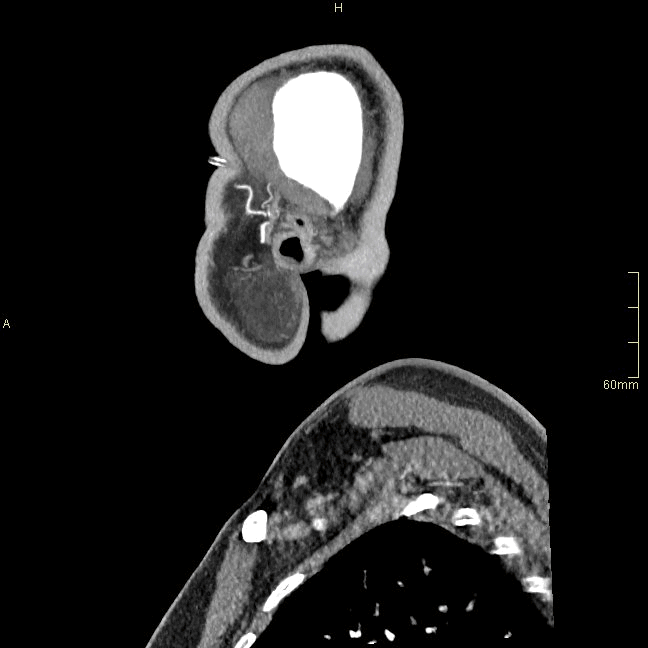
Angiography Arch to COW (Sagittal)
REFERENCES
-
Radiopaedia. CT Angiography of the Cerebral Arteries (technique). [Internet]. 2018. [Updated 01 Jul 2021, cited 29/12/2021]. Available from https://radiopaedia.org/articles/ct-angiography-of-the-cerebral-arteries-technique
-
American College of Radiology (ACR). Appropriateness Criteria. [Internet]. 2022 [Updated 2021, cited 23 Nov 2021]. Available from https://www.acr.org/Clinical-Resources/ACR-Appropriateness-Criteria
-
Anderson GB, Ashforth R, Steinke DE, Ferdinandy R, Findlay JM. CT Angiography for the detection and characterisation of carotid artery bifurcation disease. Stroke 2000; 31 (9), 2168-2194.
-
Harrison MJ, Marshall J. Indications for angiography and surgery in carotid artery disease. BMJ 1975; 1 (5958), 616-618.
-
Sullivan TM. Current indications, results, and technique of carotid angioplasty/stenting. Semin Vasc Surg 2005; 18 (2), 87-94.
-
Eisenberg RL., Johnson NM. Comprehensive Radiographic Pathology. 5th Edition. Elsevier Mosby, 2012.