This is an education resource only. Ordering of all procedure codes on this website are subject to the Canberra Health Services guidelines for imaging orders.
CT LUMBOSACRAL SPINE NON CONTRAST
NOTE: Please indicate specific vertebral levels for scan range to limit radiation if possible
INDICATIONS (1-3)
-
Primary bone tumour suspected suspected on radiography or clinical exam
-
Suspected fracture in setting of axial spondyloarthritis or ankylosis
-
Spinal tumours or vertebral metastasis
-
Suspected axial spondyloarthritis with lumbar symptoms
-
Low back pain (particular if red flags are present):
- New, history of multiple vertebral compression fractures
- New, previously treated vertebral compression fracture -
Radiculopathy, paraesthesia or myopathy, initial exam or if MRI unavailable
-
Lumbar vertebral compression fracture on x-ray:
- Asymptomatic, history of malignancy, next imaging study
- Symptomatic, new, history of malignancy, next imaging study
- Symptomatic, new, no malignancy, next imaging study -
Age ≥16 years. Blunt trauma meeting criteria for lumbar imaging. Initial imaging.
-
Pre-operative and post-operative evaluation
-
Red Flags for Spinal Imaging (this applies to all regions)
-
Sinister pain
-
Fever
-
Loss of Weight
-
Prior history of malignancy
-
Infection risk
-
Falls
-
Difficulty walking
-
PATIENT PREPARATION
-
Patient able to lie still for five minutes
-
Not claustrophobic (sedation may be given)
-
Cognitively capable of following basic instructions
-
Metal artefacts removed from the region of interest
-
No respiratory distress when lying supine
ANATOMY INCLUDED
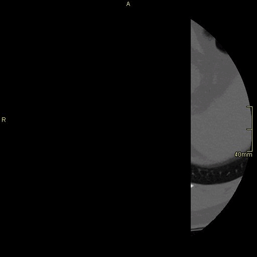
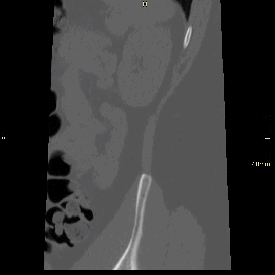

CT Lumbosacral Spine Non Contrast- Bone window (axial)
CT Lumbosacral Spine Non Contrast- Bone window (sagittal)
CT Lumbosacral Spine Non Contrast- Bone window (coronal)
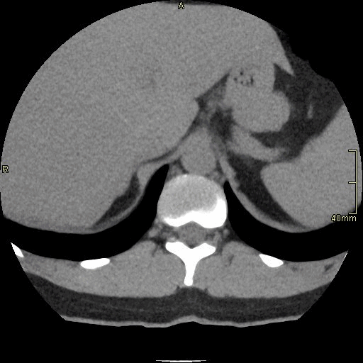
CT Lumbosacral Spine Non Contrast- Soft Tissue window (axial)
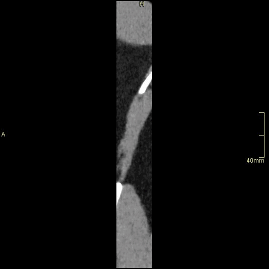
CT Lumbosacral Spine Non Contrast- Soft Tissue window (sagittal)
REFERENCES
-
Tins, B. (2010). Technical aspects of CT imaging of the spine. Insights into Imaging, 1(5–6), 349–359. https://doi.org/10.1007/s13244-010-0047-2
-
Ahmad, Z., Mobasheri, R., Das, T., Vaidya, S., Mallik, S., El-Hussainy, M., & Casey, A. (2014). How to interpret computed tomography of the lumbar spine. The Annals of The Royal College of Surgeons of England, 96(7), 502–507. https://doi.org/10.1308/rcsann.2014.96.7.502
-
American College of Radiology (ACR). Appropriateness Criteria. [ Internet]. 2022 [Updated 2023, Cited 20 April 2024]. Available from https://www.acr.org/Clinical-Resources/ACR-Appropriateness-Criteria