This is an education resource only. Ordering of all procedure codes on this website are subject to the Canberra Health Services guidelines for imaging orders.

INDICATIONS (1-5)
*This code should only be ordered after consultation with the stroke neurologist. This code is for if a Non Contrast Brain and a CT Arch to COW angio has already been performed.
IF WANTING NON CONTRAST BRAIN + PERFUSION + ARCH TO COW ANGIO-- PLEASE SEE ORDER "CT ANGIOGRAPHY ACUTE STROKE"
Potential neurological symptoms in the presence of an Acute Stroke
-
Muscular: difficulty walking, paralysis with weak muscles, problems with coordination, or paralysis of one side of the body
-
Visual: blurred vision, double vision, sudden visual loss, or temporary loss of vision in one eye
-
Speech: difficulty speaking, slurred speech, or speech loss
-
Whole body: fatigue, light-headedness, or vertigo
-
Limbs: numbness or weakness
-
Facial: muscle weakness or numbness
-
Also common: balance disorder, difficulty swallowing, headache, inability to understand, mental confusion, pins and needles or reduced sensation of touch
These symptoms should be assessed for onset and clinical correlation with a neurologists recommendation.
-
Assess whether a stroke is haemorrhage or ischaemic in nature to enact the appropriate treatment in a timely manner (e.g. endovascular clot retrieval or intravenous thrombolysis)
PATHOLOGY DEMONSTRATED (6)
Perfusion study
-
Calculation of perfusion parameters (cerebral blood flow, cerebral blood volume, time-to-peak, mean transit time)
-
Identification of penumbra vs infarct core
PATIENT PREPARATION
-
Patient able to lie still for 15 minutes
-
Not claustrophobic (sedation may be given)
-
Cognitively capable of following basic instructions
-
Metal artefacts removed from the region of interest
-
No respiratory distress when lying supine
-
Not allergic to Iodine based Contrast
-
Patient to have 18G cannula in anterior cubital fossa to allow a 7ml/s flow rate (20G cannula acceptable if flushing to 5ml/s)
-
Preferably patient fasted for 4 hours
-
Neurology to be present for scan.
ANATOMY INCLUDED
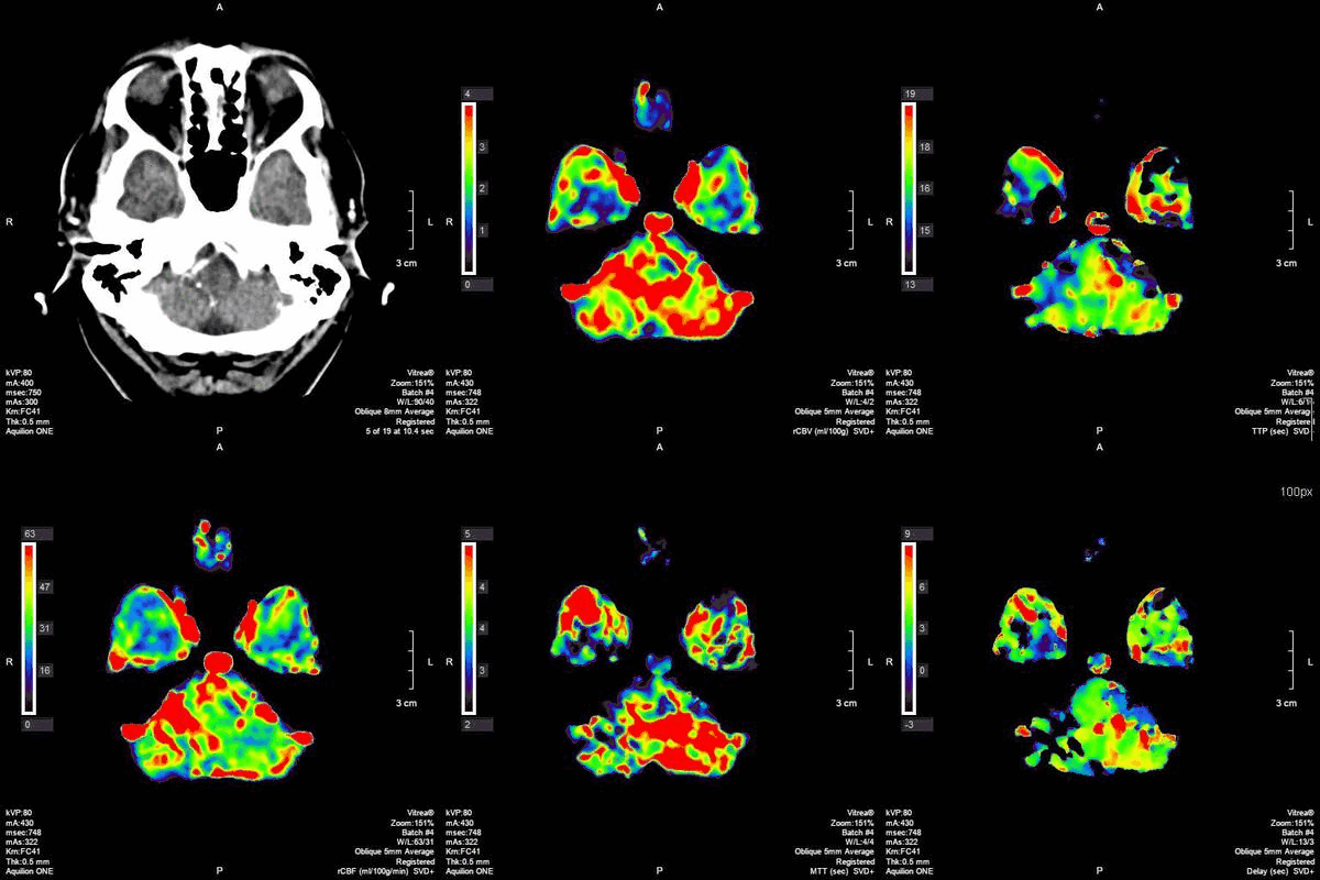
CT 4D Perfusion (Axial)
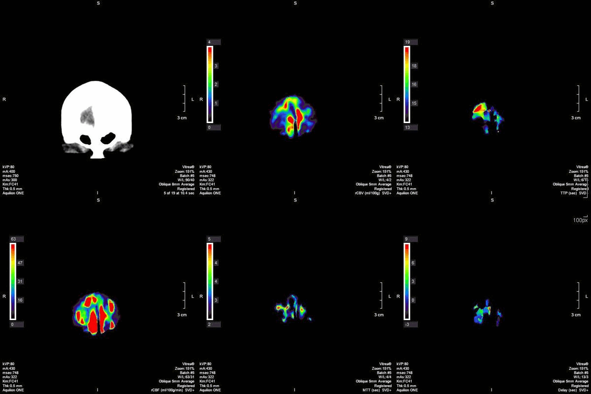
CT 4D Perfusion (Coronal)
REFERENCES
-
Radiopaedia. CT Head. [Internet]. 2010 [updated 01 Dec 2021, cited 23 Nov 2021]. Available from https://radiopaedia.org/articles/ct-head
-
American College of Radiology (ACR). Appropriateness Criteria. [Internet]. 2022 [Updated 2021, cited 23 Nov 2021]. Available from https://www.acr.org/Clinical-Resources/ACR-Appropriateness-Criteria
-
Radiopaedia. Ischaemic Stroke. [Internet]. 2011. [updated 25 Nov 2021, accessed 25 Nov 2021]. Available from https://radiopaedia.org/articles/ischaemic-stroke
-
Radiopaedia. Stroke. [Internet]. 2009. [updated 19 Apr 2021, accessed 23 Nov 2021]. Available from https://radiopaedia.org/articles/stroke?lang=gb
-
Birenbaum D, Bancroft LW, Felsberg GJ. Imaging in Acute Stroke. 2011. Western Journal of Emergency Medicine. 12 (1). 67-76.
-
Eisenberg RL., Johnson NM. Comprehensive Radiographic Pathology. 5th Edition. Elsevier Mosby, 2012